
Page 501 - Robbins Basic Pathology by Vinay Kumar, Abul K. Abbas, Jon C. Aster
P. 501
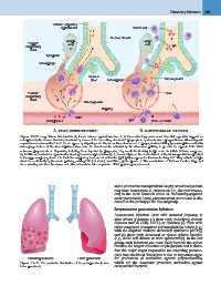
Ciliated respiratory                                           Plasma cell  Pulmonary Infections 487
                         epithelial cell
                                                                                              1
                         Lymph             1                     Mucous blanket  Lymph         IgA
                          node            2                                       node
  "Upper"
respiratory                 5

    tract

             Lymphatic

Macrophage

                                          Mucous      Macrophage                              Microorganism
                                           gland

  "Lower"               4                 Microorganism                                                     PMN
respiratory                       3                         Capillary
                                                                                              3
    tract                                                                                     Immune T cells

             Complement              PMN                                      2
                                                                       IgG

                  A. INNATE IMMUNE DEFENSES                                      B. ADAPTIVE IMMUNE DEFENSES

Figure 12–30  Lung defense mechanisms. A, Innate defenses against infection: 1, In the normal lung, removal of microbial organisms depends on
entrapment in the mucous blanket and removal by means of the mucociliary elevator; 2, phagocytosis by alveolar macrophages that can kill and degrade
organisms and remove them from the air spaces by migrating onto the mucociliary elevator; or 3, phagocytosis and killing by neutrophils recruited by
macrophage factors. 4, Serum complement may enter the alveoli and be activated by the alternative pathway to provide the opsonin C3b, which
enhances phagocytosis. 5, Organisms, including those ingested by phagocytes, may reach the draining lymph nodes to initiate immune responses.
B, Additional mechanisms operate after development of adaptive immunity. 1, Secreted IgA can block attachment of the microorganism to epithelium
in the upper respiratory tract. 2, In the lower respiratory tract, serum antibodies (IgM, IgG) are present in the alveolar lining fluid. They activate comple-
ment more efficiently by the classic pathway, yielding C3b (not shown). In addition, IgG is opsonic. 3, The accumulation of immune T cells is important
for controlling infections by viruses and other intracellular microorganisms. PMN, polymorphonuclear cell.

Bronchopneumonia                     Lobar pneumonia                   and a productive mucopurulent cough; occasional patients
                                                                       may have hemoptysis. S. pneumoniae (i.e., the pneumococ-
Figure 12–31  The anatomic distribution of bronchopneumonia and        cus) is the most common cause of community-acquired
lobar pneumonia.                                                       acute pneumonia; hence, pneumococcal pneumonia is dis-
                                                                       cussed as the prototype for this subgroup.

                                                                       Streptococcus pneumoniae Infections

                                                                       Pneumococcal infections occur with increased frequency in
                                                                       three subsets of patients: (1) those with underlying chronic
                                                                       diseases such as CHF, COPD, or diabetes; (2) those with
                                                                       either congenital or acquired immunoglobulin defects (e.g.,
                                                                       with the acquired immune deficiency syndrome [AIDS]);
                                                                       and (3) those with decreased or absent splenic function
                                                                       (e.g., sickle cell disease or after splenectomy). In the last
                                                                       group, such infections are more likely because the spleen
                                                                       contains the largest collection of phagocytes and is there-
                                                                       fore the major organ responsible for removing pneumo-
                                                                       cocci from the blood. The spleen is also an important organ
                                                                       for production of antibodies against polysaccharides,
                                                                       which are the dominant protective antibodies against
                                                                       encapsulated bacteria.